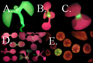
Fig. 3. Primary Screening and Characterisation(A, B, C & D). Initial screen for GFP expression in seedlings: (A) Several lines show expression throughout the seedling. (B & C) Images of seedlings from two of the lines that appear to show organ specific expression, new leaves and root respectively. (D) Vascular expression is amongst the most common components of the expression patterns we have observed, but it is often seen in conjunction with GFP expression in other tissues, e.g. the epidermis, and sometimes very specific cell types, e.g. trichomes. (E). Expanded screen for expression in the inflorescence stem, by mounting bunches of stems in wax and hand sectioning. This second screen was designed to identify patterns specific to the stem or weak GFP expression in the shoot, otherwise obscured by fluorescence from chlorophyll in the mesophyll of leaves and cortex of the stem.